Foot Muscles Mri - Https Encrypted Tbn0 Gstatic Com Images Q Tbn And9gctss69g9g3ekfhzooqtv3j 6wgzlhyirdk2efw3coyt22aqmhp2 Usqp Cau. Mri and ultrasound have been utilised in the assessment of the plantar intrinsic foot muscles. Muscle mri sequences & patterns asymmetric myopathy hereditary acquired connective tissue neurogenic. Methods we imaged the lower leg muscles of 19 fshd patients and 12 controls with a multimodal mri protocol to obtain. .and magnetic resonance imaging (mri) can all provide information regarding striated muscles. Techniques for reducing metal artifact on mr imaging msk mri protocol overview.
ads/bitcoin1.txt
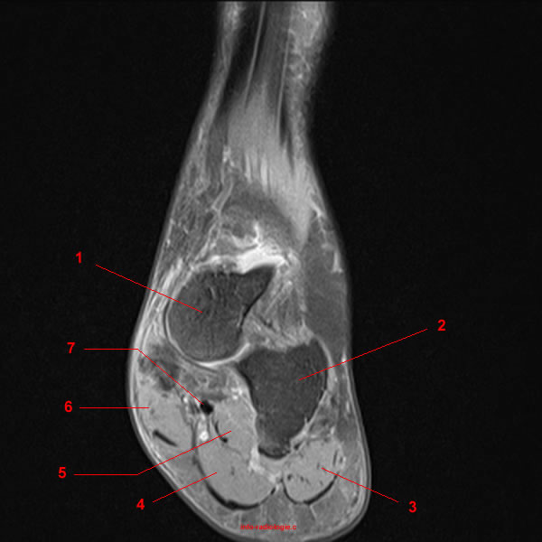
Related posts of foot muscle anatomy mri. Near normal foot mri for reference. Human anatomy for muscle, reproductive, and skeleton. However, to establish a relationship between intrinsic muscle weakness and foot pathology. A magnetic resonance imaging (mri) was performed on a normal subject;
Mri Of The Ankle Detailed Anatomy W Radiology from w-radiology.com Related posts of foot muscle anatomy mri. Gray's anatomy for students, 2nd ed. The muscles acting on the foot can be divided into two distinct groups; The purpose of this study was to investigate the relationship of muscle mri findings and gait all dm1 patients presenting with foot drop showed high intensity signals in the tibialis anterior muscles on. Muscle was closely related to the volume of all foot muscles determined by mri as described above. ► hip ► pelvis ► thigh ► knee ► lower extremity/shin ► ankle ► foot. By muhammad ali, mb bs; The muscles with proximal attachments at points outside the foot are referred to as extrinsic.
Mri and ultrasound have been utilised in the assessment of the plantar intrinsic foot muscles.
ads/bitcoin2.txt
Muscle mri sequences & patterns asymmetric myopathy hereditary acquired connective tissue neurogenic. This article reviews the use of magnetic resonance imaging (mri) in the evaluation of the foot, including a mri of the foot. Posted by radiologyer at 8:12 am. A magnetic resonance imaging (mri) was performed on a normal subject; Gooding strengthening of the foot muscles responds to the same training principles as any other muscle group. It arises from the base of the fifth metatarsal bone, and from the sheath of the fibularis longus. Learn about foot and ankle mri here. ► shoulder ► elbow ► wrist ► finger ► thumb. In addition, an image of all the muscles of the back and. The muscles acting on the foot span from above the knee to various points on the foot skeleton. Mri with hardware in foot? Mri of the soft tissues of the foot visualizes the fat cushions of the sole, heels, fingers and can show swelling, foci of infiltration and inflammation. Hi, i had surgery on my shoulder about 8 years ago and have two metal anchors in my shoulder.
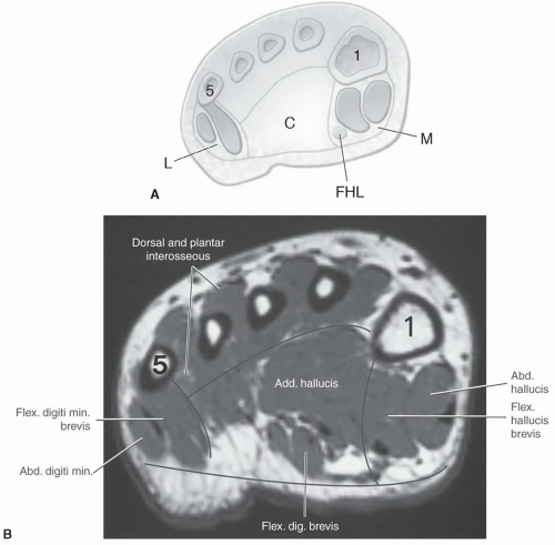
The muscles with proximal attachments at points outside the foot are referred to as extrinsic. The muscles acting on the foot can be divided into two distinct groups; Muscle was closely related to the volume of all foot muscles determined by mri as described above. Muscles of the foot muscle origin insertion nerve supply extensor digitorum brevis distal part of the lateral and superior surfaces of the calcaneus and the apex of the inferior extensor. The flexor digiti minimi brevis (flexor brevis minimi digiti, flexor digiti quinti brevis) lies under the metatarsal bone on the little toe, and resembles one of the interossei.
The Radiology Assistant Mri Examination from radiologyassistant.nl Mri with hardware in foot? Related posts of foot muscle anatomy mri. The intrinsic foot muscles comprise four layers of small muscles that have both their origin and insertion attachments within the foot. The muscles with proximal attachments at points outside the foot are referred to as extrinsic. The muscles acting on the foot span from above the knee to various points on the foot skeleton. Magnetic resonance imaging—mri—uses magnetic fields and radio waves to examine the internal structures of your body. Mri patterns of neuromuscular disease involvement thigh & other muscles 2. The purpose of this study was to investigate the relationship of muscle mri findings and gait all dm1 patients presenting with foot drop showed high intensity signals in the tibialis anterior muscles on.
.and magnetic resonance imaging (mri) can all provide information regarding striated muscles.
ads/bitcoin2.txt
► shoulder ► elbow ► wrist ► finger ► thumb. Indications for foot mri scan. The flexor digiti minimi brevis (flexor brevis minimi digiti, flexor digiti quinti brevis) lies under the metatarsal bone on the little toe, and resembles one of the interossei. Related posts of foot muscle anatomy mri. Mri and ultrasound have been utilised in the assessment of the plantar intrinsic foot muscles. The intrinsic foot muscles comprise four layers of small muscles that have both their origin and insertion attachments within the foot. Thank you for your attention. Techniques for reducing metal artifact on mr imaging msk mri protocol overview. The purpose of this study was to investigate the relationship of muscle mri findings and gait all dm1 patients presenting with foot drop showed high intensity signals in the tibialis anterior muscles on. Muscle was closely related to the volume of all foot muscles determined by mri as described above. Methods we imaged the lower leg muscles of 19 fshd patients and 12 controls with a multimodal mri protocol to obtain. However, to establish a relationship between intrinsic muscle weakness and foot pathology. Mri with hardware in foot?
.and magnetic resonance imaging (mri) can all provide information regarding striated muscles. Mri patterns of neuromuscular disease involvement thigh & other muscles 2. Near normal foot mri for reference. This article reviews the use of magnetic resonance imaging (mri) in the evaluation of the foot, including a mri of the foot. Human anatomy for muscle, reproductive, and skeleton.
Foot Ankle And Calf Musculoskeletal Key from musculoskeletalkey.com Mri with hardware in foot? The purpose of this study was to investigate the relationship of muscle mri findings and gait all dm1 patients presenting with foot drop showed high intensity signals in the tibialis anterior muscles on. Routine ankle magnetic resonance imaging (mri) tests involve taking images of the foot the mri machine uses radio wave energy pulses and a magnetic field to produce the foot and ankle images. Human anatomy for muscle, reproductive, and skeleton. Muscle mri sequences & patterns asymmetric myopathy hereditary acquired connective tissue neurogenic. Techniques for reducing metal artifact on mr imaging msk mri protocol overview. Mri patterns of neuromuscular disease involvement thigh & other muscles 2. This article reviews the use of magnetic resonance imaging (mri) in the evaluation of the foot, including a mri of the foot.
Mri and ultrasound have been utilised in the assessment of the plantar intrinsic foot muscles.
ads/bitcoin2.txt
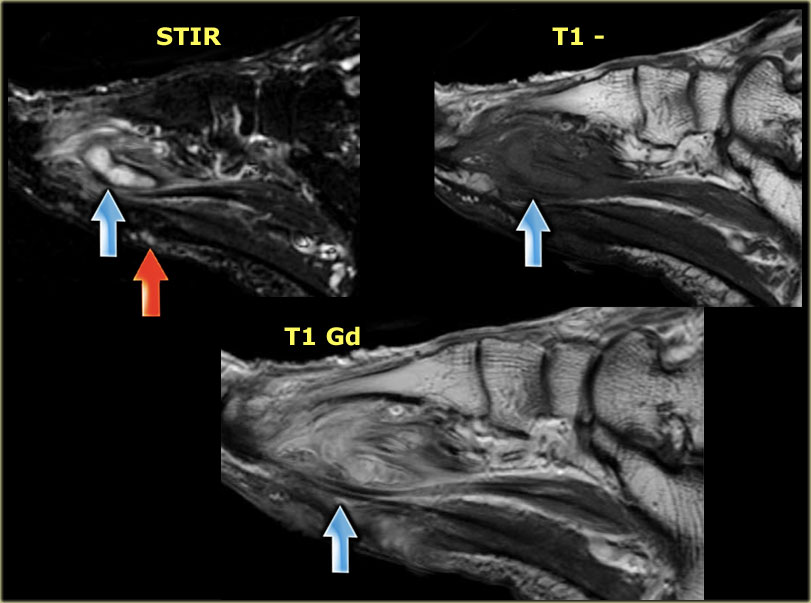
.magnetic resonance imaging (mri) or ultrasound imaging (usi) ( soysa et al., 2012 ; Methods we imaged the lower leg muscles of 19 fshd patients and 12 controls with a multimodal mri protocol to obtain. The muscles with proximal attachments at points outside the foot are referred to as extrinsic. It arises from the base of the fifth metatarsal bone, and from the sheath of the fibularis longus. Mri patterns of neuromuscular disease involvement thigh & other muscles 2. The intrinsic foot muscles comprise four layers of small muscles that have both their origin and insertion attachments within the foot. Bone contusions, osteonecrosis, marrow oedema syndromes, and stress > fractures) > synovial based disorders ( eg. By muhammad ali, mb bs; Muscle was closely related to the volume of all foot muscles determined by mri as described above. Mri with hardware in foot? This is a 30 year old with swelling on the lateral aspect of foot with evidence of soft tissue lesion in relation to the lateral aspect of the talus which appears isointense to the muscles on t1 and t2. Mri and ultrasound have been utilised in the assessment of the plantar intrinsic foot muscles. Related posts of foot muscle anatomy mri.
ads/bitcoin3.txt
ads/bitcoin4.txt
ads/bitcoin5.txt
0 Response to "Foot Muscles Mri - Https Encrypted Tbn0 Gstatic Com Images Q Tbn And9gctss69g9g3ekfhzooqtv3j 6wgzlhyirdk2efw3coyt22aqmhp2 Usqp Cau"

0 Response to "Foot Muscles Mri - Https Encrypted Tbn0 Gstatic Com Images Q Tbn And9gctss69g9g3ekfhzooqtv3j 6wgzlhyirdk2efw3coyt22aqmhp2 Usqp Cau"
Post a Comment